


EHT - the beat of advanced cardiac science
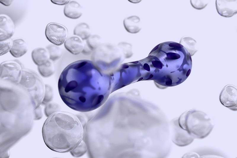
EHTs, created from human induced pluripotent stem cells, are 3D cardiac constructs that accelerate preclinical research and enhance personalized medicine studies.
Aboard the effortless journey from lab to life

We aim to make the human body measurable. One organ at a time.
Starting with the heart.
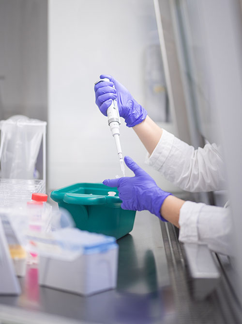
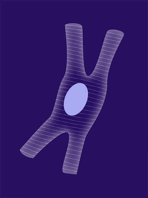
Engineer human tissue
Engineered Heart Tissues (EHTs) are three-dimensional, fibrin-based cardiac constructs derived from human induced pluripotent stem cells (hiPSCs). Our innovative technology combines hiPSCs with 3D tissue engineering to simulate human heart tissues, enabling precise cardiac safety and efficacy testing, disease modeling and precision medicine including gene therapy applications.
EHTs not only facilitate the rapid translation of research findings into preclinics but also provide a powerful testbed for personalized medicine approaches.
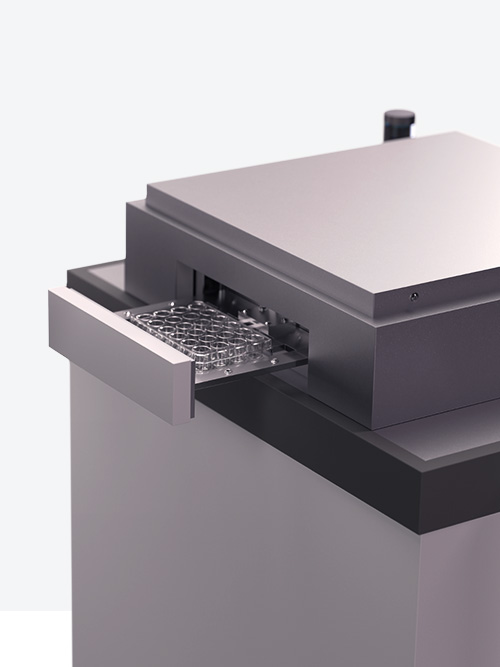
Run sterile tests in 90 s
Wouldn’t that be nice: A system that helps you discover breakthroughs with ease. That’s why we developed the DiNAQUBE™!
A fully automated EHT contraction measurement system: Its cameras capture non-invasively. It recognizes tissue with machine learning. And does it all fast: From start to result in just a minute and a half!
Now you can focus on interpretation instead of data management.
Interpret ready-to-use data
The power to quickly dive deep into your data comes with our web-based software.
It allows unprecedented comparison of multiple measurements across different plates, all in a single view. Visualized results give you clarity. And machine learning capabilities help you categorize EHTs.
All done intuitively with a few clicks. Your research should be this easy.
All benefits at a glance
Advanced technology for human heart tissue engineering
Human iPSC-derived cardiomyocytes more closely resemble human cardiac physiology than animal models.
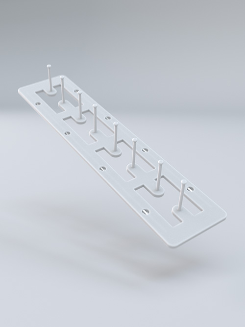
Contraction against an elastic resistance, qualitatively comparable to the load imposed on real human heart.
3D cardiac tissues reflect all compensatory mechanisms of the human heart.
Non-invasive measurements with balanced conditions.
3R compliant, human based heart model
Provides a humane alternative to animal testing in preclinical toxicology assessments
Awarded for adherence to the 3R principles of “replace, reduce, refine” in animal research*
Aligns with ethical guidelines and industry best practices, ensuring a clear conscience in research methodology
Scalable, automated solutions for streamlined research
Rapid screening with the ability to analyze 24 cardiac tissues simultaneously in 90 seconds
Increased efficiency through automated feeding and screening street
Flexibility to integrate customized secondary assays as per research needs
Access to both in-house and commercially available cardiomyocytes, ensuring comprehensive data and analysis
Efficient and intelligent data handling
Autonomous figure recognition and parameter calculation, independent of the user
Comprehensive statistics, plots and summary reports at your fingertips
Intelligent classification of contractility and tissue samples through AI
Seamless integration with big data resources for enhanced information retrieval
Consistent and reliable long-term sterile testing
Enables both short- and long-term exposure of new drug candidates
Provides a highly robust, reproducible and automated platform for controlled environment measurements with electrical stimulation options
Authentic human cardiac model for superior toxicology
Captures relevant parameters for toxicology screening, surpassing current non-human in vitro models, animal models and human heart muscle trabeculae (dying preparations)
Offers integrated functional (contractility) analyses, providing a high-content assay with the potential for additional readouts
Advanced technology for human heart tissue engineering
Human iPSC-derived cardiomyocytes more closely resemble human cardiac physiology than animal models
Contraction against an elastic resistance, qualitatively comparable to the load imposed on real human heart
3D cardiac tissues reflect all compensatory mechanisms of the human heart
Non-invasive measurements with balanced conditions
3R compliant, human based heart model
Provides a humane alternative to animal testing in preclinical toxicology assessments
Awarded for adherence to the 3R principles of “replace, reduce, refine” in animal research
Aligns with ethical guidelines and industry best practices, ensuring a clear conscience in research methodology
Scalable, automated solutions for streamlined research
Rapid screening with the ability to analyze 24 cardiac tissues simultaneously in 90 seconds
Increased efficiency through automated feeding and screening street
Flexibility to integrate customized secondary assays as per research needs
Access to both in-house and commercially available cardiomyocytes, ensuring comprehensive data and analysis
Efficient and intelligent data handling
Autonomous figure recognition and parameter calculation, independent of the user
Comprehensive statistics, plots, and summary reports at your fingertips
Intelligent classification of contractility and tissue samples through machine learning
Seamless integration with big data resources for enhanced information retrieval
Consistent and reliable long-term sterile testing
Enables both short- and long-term exposure of new drug candidates
Provides a highly robust, reproducible and automated platform for controlled environment measurements with electrical stimulation options
Authentic human cardiac model for superior toxicology
Captures relevant parameters for toxicology screening, surpassing current non-human in vitro models, animal models, and human heart muscle trabeculae (dying preparations).
Offers integrated functional (contractility) analyses, providing a high-content assay with the potential for additional readouts
Ready to research with EHT?
Your data with DiNABIOS is as secure as DNA in a double helix. Check out our Privacy Policy for the specifics.